Rapid diagnosis of head and neck lumps

Do you have a new head and neck lump?
If you’re worried about a lump that’s suddenly appeared in your neck or face, I can offer a rapid diagnostic service to give you a diagnosis, often on the same day.
What are the common causes of a neck lump?
By far the commonest cause of a neck lump is an enlarged lymph node. There are about 200 lymph nodes in the neck; they defend you from infection, injury and cancer arising from head and neck sub-sites such as skin and scalp, mouth, nasal cavities, the throat and the voice box.
Other common neck lumps include those originating in the thyroid and salivary glands (submandibular and parotid) and branchial and thyroglossal duct cysts (see below). Benign fatty lumps are called lipomas.
A laryngocoele is an uncommon, soft, neck lump which occurs when the internal lining of the voice-box protrudes through a hole in its frame-work. They are associated with high air pressure in the voice box and as such are associated with wind instrument use.
Rarer still, are benign tumours that originate from nerve cells around some of the major vessels in the neck (schwannomas and paragangliomas).
What causes enlarged lymph nodes?
Lymph nodes greater than 1 cm that persist for more than 2 weeks need further investigation; reassuringly most will be benign.
The commonest causes of lymph node enlargement are:
- Short-lived infections (viral and bacterial) that originate from the mouth (from teeth or gums for example) and throat (tonsillitis for example)
- Grumbling, prolonged infections such as TB or atypical mycobacteria (in children). HIV may present with enlarged neck lymph nodes
- Some inflammatory conditions such as sarcoidosis or systemic lupus affect the whole body, but often present with enlarged neck nodes
- Cancer metastasis. You are probably most worried about the possibility of cancer. When it does occur, the cancerous lymph nodes in the neck are usually metastasis. Metastasis are cancerous tissue that have spread from an originating site (called the primary site). Common primary sites in the head and neck include: skin and scalp, the mouth cavity, tonsils, back of tongue, the voice box, thyroid, salivary glands and the back of the nose (called the nasopharynx)
- A cancer that originates in a lymph node is called a lymphoma
How will I investigate your neck lump?
It is importance that I examine all of the potential areas in the head and neck from which a cancer can arise from. I will do this using a flexible fibre optic endoscope which will slide through your nose to give me (and you) a magnified view of the back of the nose, the throat, the back of the tongue, voice box and the upper swallowing apparatus.
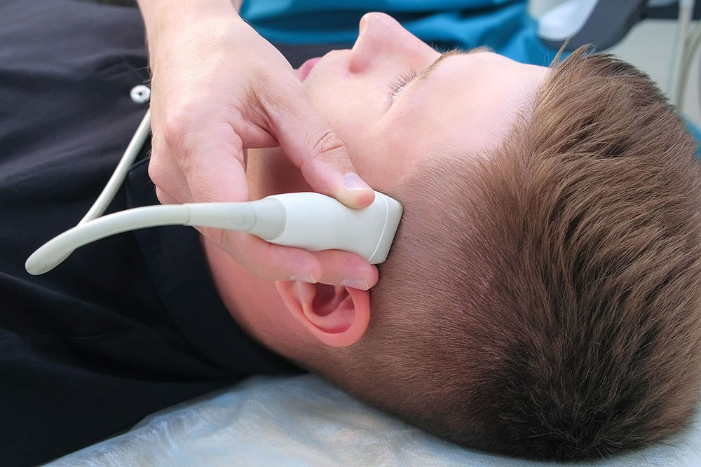
The key investigation for all neck lumps is an ultrasound scan.
I work with some of London’s leading head and neck radiologists. An experienced and skilled radiologist can distinguish between cancerous or benign lumps using an ultrasound probe. If they are concerned about a lump, they will perform a biopsy (usually a fine needle aspiration biopsy) to establish a diagnosis.
In some of my clinics, for example at OneWelbeck, an ultrasound scan can often be performed at your first visit, either just before or straight after you see me.
Specific blood tests may help me to diagnose certain benign inflammatory conditions.
I may request a CT and/ or MRI scan of the head and neck and possibly the chest if I am worried that you may have a cancerous lump.
If you do have a visible cancerous “primary” tumour on examination, it is important to try and obtain a biopsy, which may need to be performed under general anaesthesia.
2 common non-cancerous lumps that I can help you with
Thyroglossal duct cyst
This presents as a soft lump in the central part of the neck, quite high up. It moves up and down when you poke your tongue out and in respectively. A thyroglossal duct cyst is a fluid-filled collection within a piece of thyroid tissue. This thyroid tissue is left behind on a route that the embryonic, developing thyroid gland takes from its origin at the back of the tongue to its final resting place in the lower neck.
They usually present in childhood or young adulthood and can enlarge, become infected or even turn cancerous. The management is by surgical excision.
To prevent recurrence the central portion of the hyoid bone, a bone related to the base of tongue and a small portion of the muscle of the tongue base needs to be excised.
Branchial cyst
This usually presents in young adults as a soft mass on one side of the neck, just below the angle of the jaw bone. It is a skin lined ball of fluid arising in a lymph node.
In young people, they are mostly benign, but can get infected and should be treated by surgical excision.
In older people, a branchial cyst may actually be a low-grade cancer metastasis. A full head and neck examination, specialised imaging and biopsies via ultrasound are mandatory in order to exclude this possibility
