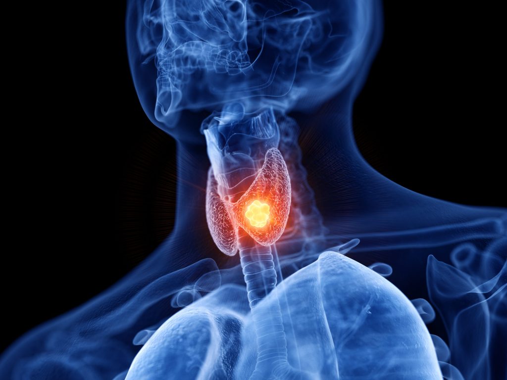
How is a Thy3 cytological diagnosis of a thyroid nodule managed?
I recommend all patients with thyroid nodules to have an ultrasound scan of the neck. If there are features that are suspicious for cancer then the radiologist will also perform an ultrasound guided fine needle aspiration (FNA) biopsy to obtain a more definitive diagnosis.
A common result from the subsequent cytology analysis is a Thy3a or Thy3f result. It can be confusing as to how to manage this scenario.
The overall risk of malignancy in patients with this result ranges from 10-40%.
Thy3a versus Thy3f
A Thy3a sample implies that there are some cellular features that are abnormal in the specimen, but does not fulfill the criteria for full blown malignancy.
If there was doubt about the adequacy of a sample, a repeat needle biopsy will usually be recommended to confirm. If it remains a Thy3a diagnosis then I would usually discuss the patient in a thyroid multidisciplinary meeting (MDT) for a consensus recommendation.
The options will be to monitor with serial ultrasound scanning (e.g. every 6 months) which will help guide a further FNA, versus a diagnostic operation to remove the half of the thyroid gland that contains the nodule (diagnostic hemi-thryoidectomy).
Features that may sway for a decision for surgery include the size of nodule (usually recommended if greater than 2 cm), if there are multiple abnormal nodules, if the patient has risk factors for thyroid malignancy (see separate article) or if the patient wants it removed because it’s interfering with swallowing, breathing or for cosmetic purposes.
A Thy3f sample, if deemed adequate, means that the lump is a follicular neoplasm. This is an overgrowth of one of the cell types making up the thyroid gland. It may be benign or malignant but a needle biopsy will not be able to distinguish between the two.
Unless the lump is less than 1 cm, or the patient is averse to an operation, the usual MDT recommendation for a Thy3f nodule would be for a diagnostic hemithryoidectomy. This is because the risk of malignancy is slightly higher in comparison to a Thy3a nodule.
If the patient does not wish to have surgery, then we would revert to serial USS monitoring.
Serial Ultrasound scanning
What we look for on serial ultrasonography is a change in the radiological features of the nodule. This may be a change in the architecture of the nodule, its size and/ or the presence of further nodules within the gland. The radiologist will also scan the remainder of the neck to check for any suspicious lymph nodes. He/ She is likely to recommend another FNA cytology if there are adverse findings.
Molecular testing
Some units perform specialist molecular tests on the needle biopsy sample to guide decision making e.g. ThyroSeq, BRAF-V600E/ RAS mutational analysis. This technology looks for mutations that are unique to thyroid cancer cells. It is important to note however, that no molecular tests as yet are 100% accurate.